Scientific Information/Data
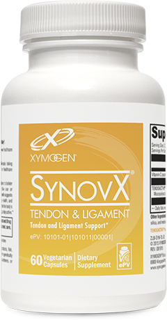
It is known that tendons and ligaments have a slower and more limited ability to self-repair than other tissues. However, healthy tendons and ligaments do indeed have an intrinsic capacity for repair, which is controlled by resident fibroblasts and their surrounding extracellular matrix (ECM).[1] Fibroblasts (e.g., tenocytes) are responsible for producing the ECM and therefore the proteoglycans (protein/mucopolysaccharide complex) and collagen needed for tissue repair. The key is to stimulate this process. In vitro and in vivo research suggests SynovX® Tendon & Ligament does just that. This proprietary blend of type I collagen and mucopolysaccharides combined with vitamin C supports the structural and functional needs of tendons and ligaments.*
Type I Collagen and Mucopolysaccharides Adult tendons are comprised mainly of type-I collagen molecules that are hierarchically organized into structural units. The molecular structure and organization of tendon and ligament collagen fibrils are key determinants in the ability of these tissues to endure mechanical force and fuel self-repair.[1] While collagen provides much of tendon/ligament structure and strength, mucopolysaccharides are said to provide the “glue” that holds them together and allows them to stretch, flex, bend, and maintain their resilience. Mucopolysaccharides—also known as glycosaminoglycans or GAGs—are a critical component of ECM and are important in maintaining structural integrity, lubrication, and spacing of collagen fibers. Furthermore, mucopolysaccharides have been shown to increase collagen and non-collagenous protein synthesis in cultures of bovine tenocytes and ligament cells.*[2]
Vitamin C This vitamin helps maintain tendon/ligament structure and biomechanical properties by stimulating collagen biosynthesis.*[3-5]
In Vitro IL-1beta (interleukin-1beta) is a cytokine associated with adverse tendon/ligament changes. The effect of SynovX® Tendon & Ligament in the presence of IL-1beta was studied in primary human tenocytes. Tenocyte cultures treated with 250, 500, and 1000 μg/ml of Synovial Tendon & Ligament showed no signs of cytotoxicity or other negative effects on the viability of cells. The major findings were that this formula counteracted the negative effects of IL-1beta by: (1) protecting tenocytes from degenerative morphological changes, cellular degeneration, and apoptosis, (2) reversing the downregulation of collagen type I and beta 1-integrin receptor expression, (3) increasing tenomodulin production, and (4) causing a significant dose- and time-dependent increase in proliferation and viability of tenocytes. These results demonstrated that SynovX® Tendon & Ligament supports tenocyte viability and proliferation and type I collagen synthesis.[1] Furthermore, the treated cells appeared healthy; displayed an abundant and well-organized ECM; and exhibited high levels of euchromatin, indicating that the cells were very active and had a high rate of protein (i.e., collagen) biosynthesis.[1] In another in vitro test, human tenocytes incubated with SynovX® Tendon & Ligament for 10 days showed a strong stimulatory effect on cell proliferation that exceeded the proliferation seen in cells incubated with (IGF-1) insulin-like growth factor 1 (positive control).[6] In addition, cells remained viable and showed large amounts of endoplasmic reticulum, which is needed for synthesis of ECM.*
In Vivo A prospective observational study performed by Nadal et al demonstrated the effects of SynoviX® Tendon & Ligament on the health of epicondyles, plantar fascia, Achilles tendons, or supraspinatus tendons. Patients were selected on the basis of clinical assessment and ultrasound results. For three months, all of the patients received 20 to 30 physical therapy sessions and the study group received two caps/d of SynovX® Tendon & Ligament.[7] Comfort, quality of life (SF-36), and physiotherapist assessments were performed before intervention began and also at 30-, 60-, and 90-day intervals during intervention. In every assessment, patients given SynovX® Tendon & Ligament showed numerical or statistically significant improvements after two to three months of supplementation compared to controls.[7] Researchers concluded that supplementing with SynovX® Tendon & Ligament improved comfort level and biomechanical properties without adverse effects.*
Several other human studies using SynovX® Tendon & Ligament have demonstrated its positive effects on tendon comfort and structure. [8-10] For instance, in a randomized placebo-controlled study (n = 60) designed to test the effects of SynovX® Tendon & Ligament versus placebo on Achilles, supraspinatus, lateral epicondyle, and plantar fascia comfort and tendon structure, Binh et al found that subjects taking SynovX® Tendon & Ligament (two caps/day) had significantly greater comfort at 90 days. At the end of the study, ultrasound assessment showed no signs of structural issues in the supplemented group.[8] In a prospective, randomized, controlled trial, 59 subjects were assigned to one of three groups: eccentric training, eccentric training plus SynovX® Tendon & Ligament, or passive stretching plus SynovX® Tendon & Ligament. Compared to physical therapy alone, the researchers found that supplementation with SynovX® Tendon & Ligament provided additional benefits associated with comfort at rest and exercise recovery as well as changes in tendon thickness and vascularization in certain subjects.*[9]
References
- Shakibaei M, Buhrmann C, Mobasheri A. Anti-inflammatory and anti-catabolic effects of TENDOACTIVE® on human tenocytes in vitro. Histol Histopathol. 2011 Sep;26(9):1173-85. [PMID: 21751149]
- Lippiello L. Collagen synthesis in tenocytes, ligament cells and chondrocytes exposed to a combination of glucosamine HCl and chondroitin sulfate. Evid Based Complement Alternat Med. 2007 Jun;4(2):219-24. [PMID: 17549239]
- Grosso G, Bei R, Mistretta A, et al. Effects of vitamin C on health: a review of evidence. Front Biosci. 2013 Jun 1;18:1017-29. [PMID: 23747864]
- Omeroğlu S, Peker T, Türközkan N, et al. High-dose vitamin C supplementation accelerates the Achilles tendon healing in healthy rats. ArchOrthop Trauma Surg. 2009 Feb;129(2):281-86. [PMID: 18309503]
- Zanoni JN, Lucas NM, Trevizan AR, et al. Histological evaluation of the periodontal ligament from aged Wistar rats supplemented with ascorbic acid. An Acad Bras Cienc. 2013 Mar;85(1):327-35. [PMID: 23460436]
- Shakibaei M, Csaki C, Mobasheri A. In vitro study of tendinopathy in humans: report on adhesion and proliferation assay, electron microscopy, immunofluorescence, and western blot analysis with Bioiberica compounds. Barcelona, Spain: Bioiberica, S.A.; 2006. [on file]
- Nadal F, Bové T, Sanchís D, et al. Effectiveness of treatment of tendinitis and plantar fasciitis by TendoactiveTM. [OARS abstract 473]. Osteoarthritis Cartilage. 2009 Sept; 17(suppl 1):S253.
- Hai Binh B, Ramirez P, Martinez-Puig D. A randomized, placebo-controlled study to evaluate efficacy and safety of a dietary supplement containing mucopolysaccharides, collagen type I and vitamin C for management of different tendinopathies. Ann Rheum Dis. 2014;73:299. doi:10.1136/annrheumdis-2014-eular.5477.
- Balius R, Alvarez G, Barb F, et al. Management of Achilles tendinopathy in reactive versus degenerative stage: a prospective, randomized, controlled trial evaluating the efficacy of a dietary supplement associated to eccentric training or passive stretching. Ann Rheum Dis. 2014;73:299-300. doi:10.1136/annrheumdis-2014-eular.5533.
- Arquer A, Garcia M, Laucirica JA, et al. The efficacy and safety of oral mucopolysaccharide, type I collagen and vitamin C treatment in tendinopathy patients. Apunts Med Esport. 2014;49(182):31-36. http://apps.elsevier.es/watermark/ctl_servlet?_f=10&pident_ articulo=90346457&pident_usuario=0&pcontactid=&pident_revista=276&ty=146&accion=L&origen=bronco%20&web=www.apunts.org&lan=en&fichero=276v49n182a90346457pdf001.pdf. Accessed January 7, 2016.


